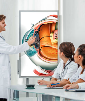
In this News Insight, ScienceRoll plots out how the field of pathology is set to evolve over the next decade. For more insights on how digital technology like AI and VR are changing healthcare, visit our healthcare solutions page. —Samsung Insights editorial team
Pathology is the motor that drives healthcare to understand diseases. While it does the job via the same methods as it did for the last 150 years, it’s time to change. Digital technologies could push the field into becoming more efficient and more scalable. They could transform the job of pathologists into a more creative and data-driven profession while allowing patients to receive diagnoses faster and more accurately. Let’s see how the digital future of pathology looks!
The foundation of medicine, pathology, has not changed for over 150 years
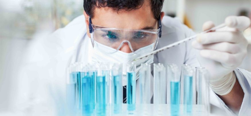
Although the whole edifice of medicine rests on the pathologist’s diagnosis, the field has not experienced any significant change for the last 150 years; said Thomas Fuchs, Director of Computational Pathology Lab at Memorial Sloan Kettering Cancer Center and Founder of Paige.AI, which is building machine learning algorithms to help digitize pathology, at the NVIDIA GPU Tech Conference in March in San Jose. That basically means that in spite of the rapid development of medical technology, such as blood-drawing robots, exoskeletons, augmented reality, synthetic organ tissues, the core of the diagnostic process is still resting on pathologists looking at tissues with a microscope in a lab. Approximately 90 percent of the work is yet done on various stains with methodologies that doctors came up more than a hundred years ago.
Moreover, the entire process of analyzing the stains is highly subjective and could signify a cumbersome process. For example, in the case of a prostate biopsy, pathologists have to scrutinize around 48 slides. Also, the results depend on the specialty, the personal judgment and more often than not even on the mental state of the pathologist – lowering the levels of accuracy.
In addition, it is not easy to compare cases. Pathologists are doing enormous work and spend too much time on searching for old slides, images, and case information in the archives of hospitals. While retrospective data use is possible in pathology – Fuchs mentioned that there are perfectly utilizable slides even from the 1930s – this amazing resource cannot be leveraged on efficiently enough as you have to look up decades-old tumor slides manually. In the 21st century, the age of digital assistants making appointments in restaurants or hair salons, it is painful to even hear about that.
Pathologists are scarce assets
No surprise that pathologists’ are “limited assets” worldwide, and their number is predicted to decrease worldwide. According to one study, by 2030 the number of active pathologists may drop by 30 percent compared to 2010 levels. In some parts of the globe, the lack of pathologists is shocking. According to the Chinese Pathologist Association, there are only 20,000 licensed pathologists in the entire country with a population of over 1.4 billion people. The situation is even worse in some parts of Africa and Asia. Malaysia has one pathologist for every 100,000 patient, while this number is sevenfold in Sudan and even worse in Uganda. There, the job is basically non-existent: the statistics enumerate 1,555,000 patients per pathologist.
The situation is not rosy in the OECD countries either. In the United States, practicing pathologists will begin to retire at an increasing rate, peaking in 2021, far faster than the current replacement rate. At the same time, the number of cases is increasing, and pathologists are forced to take on more and more.

Source: www.clpmag.com
How could digital technology change the status quo?
The digitization of pathology labs makes the specialty more efficient, the specimen more reproducible, the work of pathologists less cumbersome. The digital whole-slide imaging (WSI) allows the capture and visualization of the entire tissue on a slide, as opposed to the narrow field of view that a microscope provides. It could make pathology more accurate and reduce subjectivity.
Moreover, a digitized slide could spare enormous amount of time for physicians. Within the hospital, it often happens that various tissues are delivered from the lab to the surgeon than back again to the pathologist. Sending over a digitized slide is both safer and faster. Moreover, it allows computerized quantitative analytics, speedier image search, and slide retrieval when it comes to old cases. It reduces the barriers between hospitals, regions, and countries facilitating consultations and referrals. Not to speak about the possibility for easing the burden caused by the lack of pathologists worldwide. Just imagine how many Chinese hospitals could benefit from digitized pathology labs when they could share their slides in a cloud and retrieve all the information from it. Of course, we are not looking at the accessibility, the privacy issues or the political feasibility of such an experiment here; those are other topics.
The use of digital technologies, first of all, A.I. could help make the field “more robust, more producible, and it could really push the field from a purely qualitative to a quantitative area,” noted Fuchs in an interview with The Medical Futurist. So, let’s see the array of new technologies which could support pathology’s bright future!

Source: jpathology.com
1) Digitized workflow – from electronic microscopes to the cloud
The role of the pathology laboratory is to use blood, fluid or tissue to provide accurate, timely and relevant information useful in making diagnoses, and to guide and monitor therapy. After macroscopic evaluation of the masses in the digitized pathology lab, the process of whole slide imaging takes place. After the slices are prepared, an electronic microscope digitalizes the samples for further analysis with the help of various scanning solutions. A variety of digital products are already available on the market for migrating the entire workflow of pathologists from the manual to the digital. Whole-slide imaging solutions, such as the Nanozoomer, scanners, software, storage systems, clouds and communication systems work together to ease the work in the labs.
It will make sending slides and data for consultation much easier so providing virtual pathology services at remote sites and facilitating discussions and second opinions will undoubtedly become a booming area. Although we will always have someone looking at tissue through an electronic microscope – the interpretative reporting will be carried out in front of pulsating screens rather than next to microscopes.
However, the digitized workflow also comes with limitations that need to be solved in the future for a seamless operation. As pathology images require very high resolution and image quality is key, files have enormous sizes. Storage of huge datasets remains a challenge as well as their transport through various computer systems.

Source: https://www.youtube.com/watch?v=lp_c-hlS_w8
2) Deep learning for automation
The digitization of pathology resources opens the door for the utilization of the massive potential of artificial intelligence in the field. However, the first step for that is digitization. Fuchs says that they have been digitizing slides for the last five years at Memorial Sloan Kettering – 40,000 slides per month. Moreover, with Paige, they are speeding up the process for at least 200,000 slides, but it will still take up years until the system will be extended enough for clinical use. In fact, he added, “no one wants to use algorithms trained on small data sets, that would be like training your self-driving car in a parking lot.”
Nevertheless, once the data sets are large enough, deep learning algorithms will be for pathologists as if they have hit the bonus.
They could be used for the classification of digitized pathology slides – similarly to the practice used in radiology. Just as Facebook recognizes faces in photos, these image recognition systems identify slides with precancerous or cancerous cells, and groups them accordingly. Fuchs mentioned that, interestingly enough, when having worked for NASA, they used similar algorithms to detect volcanoes on Venus as for discovering cell nuclei.
Anyhow, the process enables pathologists to spend more times on cases requiring more attention and to set aside simpler slides with benign tumors. Thus, they will have more time on their hands, which might bring another solution for the lack of pathologists worldwide.
3) AI for better augmentation, prognosis, and prediction
Pathologists use many techniques for augmenting tissues and samples to make specific abnormal mutations visible. For example, molecular tests have been used for many years to illuminate proteins. Deep learning algorithms will be able to do something similar with digitized images in the future. A.I. could spot certain linkages, which are not visible for the human eye.
For example, augmentation could integrate computer-quantified histology data from a slide with data from other sources, such as DNA sequencing, helping pathologists to conclude with higher accuracy and confidence. Another use of augmentation is counting and differentiating cell types to help with the staging of tumors.
These methods also give more room for prognosis and predictions. The latter is the most positive addition of deep learning algorithms. Fuchs says that in the future when you have a three-cent H&E slide, an A.I. algorithm could tell you there is an indication caused by a specific mutation. That could lead the specialist to sequence precisely that, and come up with a targeted treatment for the patient.
When formulating a diagnosis, algorithms could be of tremendous help. A study showed that the combination of a deep learning system’s predictions with the human pathologist’s diagnoses for identifying metastatic breast cancer resulted in 85 percent reduction in human error rate! That is simply amazing!

Source: www.hbr.org
4) Biotechnology for precision medicine
Various digital technologies will improve the point-of-care testing (PoCT) options of pathology departments in the future. Just as portable diagnostic devices are making it possible to diagnose patients whenever they are, such tools enable the examination of tissues, fluids and other samples near the patient’s location, in the surgery or clinic at the time of consultation to facilitate prompt clinical decision making regarding patient management. Various blood glucose meters, urine test strips, pregnancy tests fall into the category of PoCT, but their range will significantly widen in the future.
Some start-ups are already raising the stakes. The UK-based start-up, Oxford Nanopore Technologies offer real-time, out-of-the-lab DNA and RNA sequencing with their MinION device. It enabled researchers to carry out offline DNA sequencing of environmental samples on Antarctica using only a hand-held device. Mind-blowing, isn’t it?
Molecular analysis will enable more precise diagnosis in the future, and revolutionize how we define several diseases. It will also give precious information on how patients respond to treatment and what is the prognosis. The pathologist will help interpret the results and choose which molecules to be evaluated.

Source: barnsleyhospital.nhs.uk
5) Pathology and radiology
There are several similarities between the two medical fields. They are both necessary for the diagnostic process, and they both represent a qualitative science based on the opinion of medical experts. Lately, there are also common features how they are digitized. Pathologists and radiologists both look at anatomies through imaging devices – although one of them at the cellular level, the other at the organ level. Moreover, due to the rapid changes in digital imaging methods in the last years, both fields are transforming faster than any other medical specialties.
Fuchs explained that pathology will be fundamentally different in 10 years, but he does not believe that, for example, the fears of A.I. taking jobs resonate the same way in pathology as it does in radiology. He thinks that radiology, dermoscopy, and other disciplines might be more scared because these are rather screening steps. However, as soon as you see something in radiology or dermoscopy, you have to look at the pathology. So that’s the ground truth. After all, everything is based on that diagnosis. Thus, the human will always be there, looking at the microscope. No matter whether there is an A.I. or any other technology collaborating with it.

Source: xiahepublishing.com
Pathology is heading towards the cosmos
However, all the above-enumerated factors will definitely change the specialty – but Fuchs believes it is going to be more exciting for pathologists. The Medical Futurist agrees. If imaging methods are powerful enough to analyze individual cells, pathology could become an entirely different branch of medical sciences. A new chapter could start for traditional pathology that goes down to the level of individual cells instead of looking at the organism as a whole. At this point, radiology and pathology might even overlap/merge to some degree as one will have to know both areas.
Both physicians and patients will benefit from this transformation. With better tools, medical professionals will have more chance to save lives and patients will have more chance to survive. With pathology, it’s a little bit like cosmology. When astronomers look for exoplanets that we don’t yet fully understand/know, the better telescopes they can use, the more we will learn.
The views expressed in content distributed by Newstex and its re-distributors (collectively, “Newstex Authoritative Content”) are solely those of the respective author(s) and not necessarily the views of Newstex et al. It is provided as general information only on an “AS IS” basis, without warranties and conferring no rights, which should not be relied upon as professional advice. Newstex et al. make no claims, promises or guarantees regarding its accuracy or completeness, nor as to the quality of the opinions and commentary contained therein.
This article was written by nora from ScienceRoll and was legally licensed through the NewsCred publisher network. Please direct all licensing questions to legal@newscred.com.
Learn how new collaboration technologies can help healthcare practitioners and researchers work smarter.
![]()